


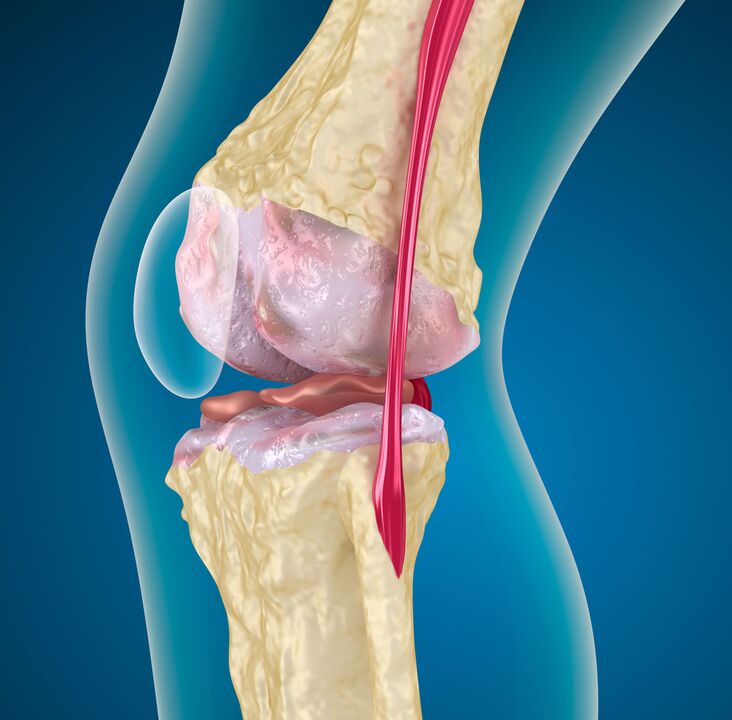
Up to 20% of all people over the age of 25 are at risk of developing osteoarthritis deformans of the knee. The knee joint works in axial compression mode, therefore, its articular surfaces are subjected to constant loads and are subject to degenerative changes in the hyaline cartilage.
%20and%20affected%20by%20osteoarthritis%20(left).jpg)
Prevalence
Pathology of articular cartilage of a degenerative-dystrophic nature with the involvement of bone tissue, joint bag, ligaments and muscles in the process is called deforming arthrosis. In terminology there are synonyms:
- arthrosis;
- arthrosis;
- degenerative arthritis;
- arthrosis;
- hypertrophic arthritis;
In terms of frequency, damage to the knee comes immediately after the hip joint, so a stable phrase has formed - "gonarthrosis of the knee joint". The dependence of the frequency of the disease on age was studied:
| 26 - 44 years old | 5% of adults |
| 45 - 59 years old | 16. 70% |
| 60 - 69 years old | 12. 10% |
| 70 years and over | eleven% |
In all age groups, the representatives of the fair sex quantitatively predominate. In them, knee osteoarthritis occurs 1. 2-1. 4 times more often than in men.
In the field of persistent disability, deforming osteoarthritis of the knee joint accounts for nearly 30% of all causes of disability associated with joint pathology.
Classification of gonarthrosis
For the sake of development, the disease is divided into two large groups: primary and secondary. The primary arises without visible prerequisites. The secondary is preceded (or accompanied by) provoking factors:
- biomechanical disorders: trauma, excessive loads, developmental abnormalities (dysplasia), skeletal pathologies (scoliosis, flat feet), obesity;
- inflammatory processes: aseptic or infectious arthritis, hemarthrosis frequent in hemophilia;
- metabolic diseases: gout, hemochromatosis, Paget's disease;
- endocrine gland disorders: acromegaly, diabetes mellitus, parathyroid gland disorders;
- violations of adequate blood supply: varicose veins and post-thrombophlebitic syndrome, obliterating endarteritis, atherosclerosis of the vessels of the lower extremity;
In medical practice, classification according to the severity of pathological changes is more useful. The evaluation is made on the basis of radiographic studies. The most popular clinical and radiological classification.
I stage
The picture shows a slight narrowing of the interarticular gap (comparison is made with a healthy joint), the onset of sclerosis of the pericartilaginous bone tissue. Clinically: Pain occurs during walking or immediately after, with prolonged standing. More pronounced when climbing stairs. Go to rest. 1st degree gonarthrosis does not greatly affect the function of the joint.
II stage
The joint space is 2-3 times narrower than normal. Sclerosis is more pronounced, osteophytes are found (spiky growths of bone tissue along the edges of the joint space and condyles). The pain is moderate, there are signs of muscle hypotrophy, lameness. Deformation of the knee in the frontal axis is visible. 2nd degree gonarthrosis leads to a significant limitation of joint mobility.
III stage
Sclerosis of cartilaginous elements, deformation of the articular surfaces. There are areas of subchondral necrosis, local osteoporosis. Cyst in the adjacent bone tissue. The joint space is critically narrowed, sometimes not defined. Large osteophytes. Atrophy of the muscles of the thigh and lower leg, the joint is unstable, there is a pronounced deformity. Movement in the knee is sometimes impossible, contracture is formed. When moving - severe pain, lameness.
This approach to classification is convenient in that it allows you to assess clinical manifestations in relation to organic changes. It offers the opportunity to choose a more effective treatment based on a comprehensive assessment of the state of the joint.
Development mechanism
The pathogenesis of any arthrosis goes through three stages:
- Damage to cartilage microstructures. Under the influence of any of the damaging factors, compounds with a high molecular weight lose their strength and are enriched with water molecules. The ability of low molecular weight collagens to assemble into macromolecules is impaired. This leads to a decrease in the strength and durability of the hyaline cartilage. Chondroprotectors counteract these phenomena.
- If the provoking factor is not eliminated, the weakening of cartilage components (glycosaminoglycans, proteoglycans) continues. This leads to the activation of recovery processes. Their power reserve isn't particularly large, so this stage quickly transitions into the next.
- The disruption of the compensatory mechanisms leads to the progressive destruction of the articular cartilage, the death of its cells - chondrocytes. Cartilage cracks extend to the underlying bone. The degree of detachment of cartilaginous components increases, their defibration occurs, which leads to thinning of the hyaline membrane.
On the part of the bone, with deforming arthrosis of the knee joint, thickening (sclerosis) occurs, cysts appear and areas with uneven bone density. Thus, deformation of the articular surfaces develops, instability of the joint.
Diagnostics
The diagnosis is based on a set of data obtained as a result of a survey (anamnesis), medical examination and instrumental research methods. The latter include radiographic examinations (CT, MRI), radioisotopes (scintigraphy), arthroscopy.
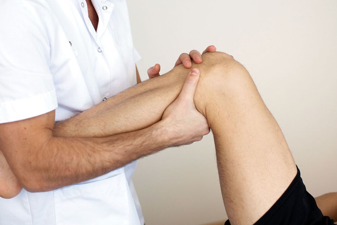
Objective examination
It includes clarification of the patient's life history, the circumstances preceding the development of gonarthrosis of the knee joint, collection of complaints and examination. In the process, the presence of provoking factors and the degree of their influence on the development of the disease are clarified.
At this stage, it is important to find out the condition of the second knee. If you miss bilateral knee OA and focus only on the knee that worries you the most, you may be making a gross diagnostic error.
For this, functional tests should be performed on two limbs simultaneously. Attention is drawn to the pain of active and passive movements, sensitivity on palpation, crepitus (crunch) during extension and flexion. Chronic inflammatory processes lead to the appearance of a Becker's cyst - a protrusion of the joint sac in the popliteal fossa, which can also be detected by palpation.
instrumental methods
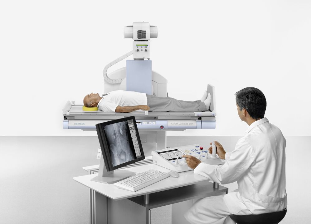
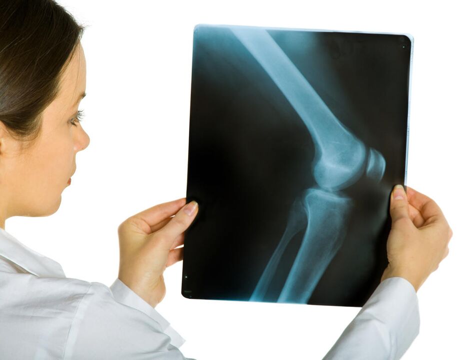
The first is the radiography. A two-view image of the knee allows a preliminary assessment of the condition of the joint and determines the stage of the disease. The disadvantage of the method is that radiological signs occur later than the symptoms and morphological changes accompanying arthrosis of the knee joint.
In these cases, MRI (MRI) helps. It is possible to determine the initial stages of degenerative changes in cartilage and bone structures, it is possible to assess the state of the intra-articular ligaments, menisci. Scintigraphy for gonarthrosis of the knee joint provides data on the functional status.
Direct examination of the joint cavity is possible with arthroscopy.
For the unification of diagnostic data, the American College of Rheumatology has proposed the following criteria:
- Age over 50 years.
- Joint stiffness in the morning, which persists for at least half an hour.
- Cracking, determined by movement (active and passive).
If these symptoms are accompanied by osteophytes detected on x-rays and pain, gonarthrosis of the knee joint is very likely to occur.
The initial stages of the disease may not be pronounced, therefore it is necessary to carry out a differential diagnosis with other joint pathologies, in which pathogenetic drugs for osteoarthritis (chondroprotectors) will be ineffective.
All possible measures must be taken not to confuse gonarthrosis with the following conditions:
Rheumatoid arthritis |
Early age onset, stiffness in the morning for more than 30 minutes, pain worse at rest and weaker on movement, rheumatoid nodules on skin, concomitant lesions of internal organs, symptoms of intoxication (fever, sweating), C-reactive protein in tests some blood. |
Crystal Arthritis |
The pain is sharp, at night or in the morning; the skin over the diseased joint is swollen, red, hot; crystals on a microscopic examination of the synovial fluid, an increase in uric acid in the blood (with gout). |
Spondyloarthropathies |
Arthritis of other unrelated joints (intercostal joint, lumbar); inflammatory processes in the tendons; damage to the cornea, skin, mucous membranes. |
In the International Classification of Diseases of the 10th revision (ICD 10), all these diseases are assigned the index "M", but a different numerical code.
These are fundamentally different pathological processes that require a professional approach to diagnosis and qualified treatment.
Therapeutic measures
If there is a disease, there must be ways to cure osteoarthritis of the knee joint. And they exist. Help can be provided in a variety of ways.
First there are the achievements of traditional medicine, based on a thorough study of the causes and mechanism of the disease. Here, medical and surgical methods are used. Competent treatment requires consistent and complex use of drugs, methods of physiotherapy and rehabilitation measures.
The second way is treatment with folk remedies. The effectiveness of these methods, to put it mildly, is questionable. But they are used because it is possible to reduce the manifestations of the disease at home. Folk remedies can be used only as an addition to drug treatment or as part of complex therapy, it is imperative to obtain the consent of the attending physician!
medicinal aid
This type of treatment includes the use of various medications. For medicinal effects, drugs of several groups are used:
- non-steroidal anti-inflammatory drugs, analgesics, opiates;
- slow-acting symptomatic drugs (chondroprotectors);
- glucocorticoid hormones;
NSAIDs, rapid analgesics, opiates
Medicines of this group are designed to eliminate pain. Pain syndrome practically spoils the life of patients with arthrosis, its removal significantly improves the quality of human life. NSAIDs, anilides, non-narcotic and narcotic analgesics are capable of this.
A common drawback is side effects. These drugs negatively affect the kidneys, the protective mechanisms of the gastrointestinal tract. An alternative that can reduce the harmful manifestations are injections. Intramuscular administration harms the stomach less and accelerates the effect.
Due to side effects, drugs of this group are prescribed during exacerbations, careful selection of the dose is necessary.
The main advantage of NSAIDs is the numerous forms for local treatment (ointments, gels). It allows you to control the manifestations of the disease at home.
Centrally acting analgesics are prescribed for a short time, with the other two groups ineffective. The most popular opium is prescribed during an exacerbation, most often with bilateral gonarthrosis. These drugs are addictive. You can't get them yourself!
Slow-acting symptomatic drugs

The action of these substances is twofold: they have the ability to reduce pain (like NSAIDs) and contribute to the restoration of hyaline cartilage. They are often called chondroprotectors.
The effect develops over several weeks (2-8) and persists after cancellation for 2-3 months.
In addition to chondroitin sulfate and glycosaminoglycans, this group includes preparations based on hyaluronic acid, compounds derived from avocados and soybeans.
The most studied and popular chondroprotectors (chondroitin sulfate and glycosaminoglycans) are ready-made components of articular cartilage. Well absorbed into the blood, forms high concentrations in the joint cavity. To speed up the effect, injections can be made directly into the joint.
Chondroitin sulfate, taken in cycles for two years at a daily dose of 800 mg, has been shown to have a stabilizing effect on the joint space in gonarthrosis of the knee joint of the 2nd degree.
Avocado/soy compounds have anti-inflammatory effects. Due to the inhibition of collagenase (a decaying enzyme), they significantly slow down the destruction of cartilage, increase the synthesis of "their own" collagen. They are also very well tolerated.
Hyaluronic acid derivatives are used in the form of intra-articular injections. These funds, like chondroprotectors, improve the functional state of the knee joint.
The mechanism of action of various slowly symptomatic drugs is slightly different, therefore their combined use is recommended. A high level of safety allows you to take chondroprotectors for a long time without tangible harm to the body.
Glucocorticosteroids
The main action is anti-inflammatory. These funds are prescribed when NSAIDs are ineffective. Tablet forms also damage the stomach lining. There are forms for intra-articular injections.
They have numerous side reactions, so you should not abuse hormonal drugs to deform arthrosis of the knee joint.
Group name |
Benefits |
Cracking |
|---|---|---|
NSAIDs, analgesics, opiates |
Fast acting, many forms for topical application. |
Side effects, unstable effect, dangerous for age-related patients, addiction occurs. |
Chondroprotectors |
They act pathogenetically, have a lasting effect, are non-toxic, there are forms for external and intra-articular use. |
Slow development of effect. |
Hormones |
Quick effect where NSAIDs are not enough; forms for intraarticular administration. |
Side effects, unstable effect, long-term use is impossible. |
ethnoscience
At home, you can reduce the manifestations of the disease with folk remedies. There are many recipes, but there are some but:
- no clinical studies have been conducted;
- it is impossible to accurately dose the medicinal substance;
- indications are not clearly defined;
- individual tolerance of folk remedies is not taken into account;
Benefits include a broad therapeutic range, a large selection for external use. Homemade compresses and tinctures, ointments are popular.
The effectiveness of home treatment can be confirmed by the fact that biologically active substances (gum, bile, infusions of medicinal plants) are used for the preparation.
Also, competent treatment with folk remedies begins with adherence to a diet, weight loss. This method alone, aimed at reducing the load on the joint, can reverse arthrosis of the knee joint of the 1st degree (the condition is young age, sufficient compensatory capabilities). A healthy diet, in itself, stimulates the regenerative capacities of the body. The diet includes: a slight feeling of hunger, greens, freshly squeezed juices. It is recommended to add low-fat jellies, jellies to the diet.
External means are very diverse. They mainly have an irritating and warming effect. The most studied components are bile, dimethyl sulfoxide and bischofite. Bile should be used medicinally and not independently extracted from an animal's corpse. Dimethyl sulfoxide is an analogue of a chemical warfare agent, mustard gas. Bischofite is a petroleum derivative. This is the difference of origin.
All three drugs have an anti-inflammatory effect, however, they should be used at home only after consulting a doctor. These substances also have contraindications and application features.
We must not forget about the placebo effect in the treatment of folk remedies.
The last thing I want to convey is that a person has health. You should not fully rely on the seeming simplicity and cheapness of folk remedies. If you've already decided to try them, increase your focus on the sore joint. The progression of the disease against the background of treatment with folk remedies is a reason to reconsider the approach to therapy.
If osteoarthritis of the knee joint grade 2 or higher is diagnosed, it is better not to mess with traditional medicine. Or postpone it for a period of remission. Unsatisfactory treatment is an indication for a complex surgical intervention.